Electromyography (EMG) is a diagnostic procedure to assess the health of muscles and the nerve cells that control them (motor neurons). EMG results can reveal nerve dysfunction, muscle dysfunction or problems with nerve-to-muscle signal transmission. Motor neurons transmit electrical signals that cause muscles to contract. An EMG uses tiny devices called electrodes to translate these signals into graphs, sounds or numerical values that are then interpreted by a specialist.
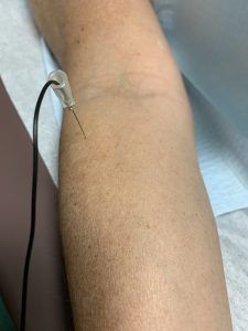
During a needle EMG, a needle electrode inserted directly into a muscle records the electrical activity in that muscle.
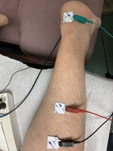
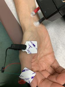
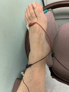
A nerve conduction study, another part of an EMG, uses electrode stickers applied to the skin (surface electrodes) to measure the speed and strength of signals travelin between two or more points.
Why it’s done
Your doctor may order an EMG if you have signs or symptoms that may indicate a nerve or muscle disorder. Such symptoms may include:
- Tingling
- Numbness
- Muscle weakness
- Muscle pain or cramping
- Certain types of limb pain
EMG results are often necessary to help diagnose or rule out a number of conditions such as:
- Muscle disorders, such as muscular dystrophy or polymyositis
- Diseases affecting the connection between the nerve and the muscle, such as
myasthenia gravis - Disorders of nerves outside the spinal cord (peripheral nerves), such as carpal
tunnel syndrome or peripheral neuropathies - Disorders that affect the motor neurons in the brain or spinal cord, such as
amyotrophic lateral sclerosis or polio - Disorders that affect the nerve root, such as a herniated disk in the spine resulting
in radiculopathy
NCV – Nerve Conduction Velocity
Small metal electrodes are taped to the skin and the nerve is stimulated by a small electric impulse. This lasts a very short time, such as 1/1000 of a second. The stimulus is slightly stronger than the electric static shock from touching a door knob after walking across a carpet.
EMG – Electromyography
If the nerve to a muscle is damaged, the muscle shows abnormal electrical activity. A few muscle diseases as well
can be detected by this test. A small Teflon coated wire pin electrode is placed in the muscle to record the activity.
There is minimal discomfort with the procedure. Because the wire is so thin, there is almost no risk of bleeding or
infection.
How should I prepare?
These tests can be performed under most any medical condition.
Please have your skin free of dirt, creams and oils.
Do not apply lotion before the test.
On cold days, it is best to arrive early. It is better if the tested area is warm. If possible, wear casual clothing, as you
may need to change into a gown before the test.
If you are unable to make it to your test on time, please call. The test can always be re-scheduled. The test generally
takes 15-45 minutes, but it may be difficult to start the test late if another patient is scheduled immediately afterward.
How will I pay for my test?
This test is routine and is covered by any standard insurance. A small number of insurance plans require a referral
from your family doctor. Please check referral requirements with your insurer or with the person who schedules your
test.
When will I learn my results?
Your doctor will discuss your results on your return visit. This test is only one piece of information which he/she will
need to put together with the other details of your case.
Please Note…
Only the doctor and the patient will be in the testing room. Please do not bring small children who cannot wait alone
in the waiting room. The patient will return to the waiting area at the completion of the test.